Polarization and functional specialization of cells

Cell polarity – the asymmetric distribution of components and functions within a cell along a directional axis – is a fundamental property of cells that is present across the kingdoms of life. Most cells need to distribute proteins, organelles, and functions along an axis of polarity in order carry out their specialized functions and organize the 3D multicellular body. We use a combination of systems biology approaches, systematic experimental manipulation, and live-cell imaging to study how cells polarize, specialize, and become organized into functional tissues.
The Boxem group is part of the Institute of Biodynamics and Biocomplexity at Utrecht University. Utrecht University is a friendly and ambitious university at the heart of an ancient city. We love to welcome new scientists to our city – a thriving cultural hub that is consistently rated as one of the world’s happiest cities. The Boxem group works closely together with the groups of Dr. Martin Harterink (also a member of NERVSPAN), Prof. Sander van den Heuvel, and Dr. Suzan Ruijtenberg who also primarily use C. elegans as a model system. You will therefore be part of an interdisciplinary and dynamic team of researchers that share many common interests and approaches.
Learn more on our team's website here.
Contact Mike BOXEM at m.boxem@uu.nl
NERVSPAN project
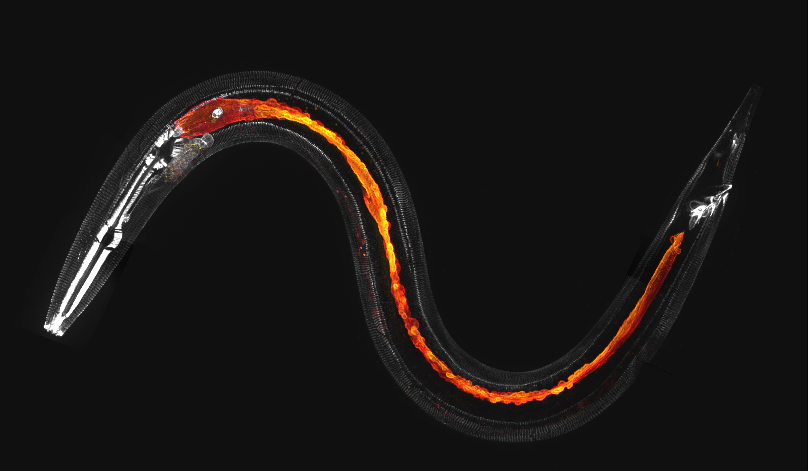
The aim of this project is to identify and characterize novel regulators of cell polarity. Two of the most abundant polarized cell types of the animal body are epithelial cells and neurons. Epithelial cells establish functionally distinct apical and basolateral domains to form selectively permeable barriers, while neurons form molecularly distinct axons and dendrites to enable directional information flow.
Objectives
Your project will involve:- the identification of novel regulators of cell polarity through protein interaction mapping approaches (proximity labelling) and genetic screens;
- validation of candidate components in vivo through localization studies (live cell imaging using endogenously tagging with fluorescent proteins);
- detailed functional analysis of validated candidates in neuronal and epithelial tissues.
In your studies you will use the nematode C. elegans as a model system. Within this animal model, the polarization of cells can be followed with single cell resolution, and polarity regulators can be studied using the advanced genetic toolkit available, including CRISPR/Cas9 genome engineering to generate mutants and inducible degradation variants, to be able to inactivate candidates in specific cells and at specific times in development.